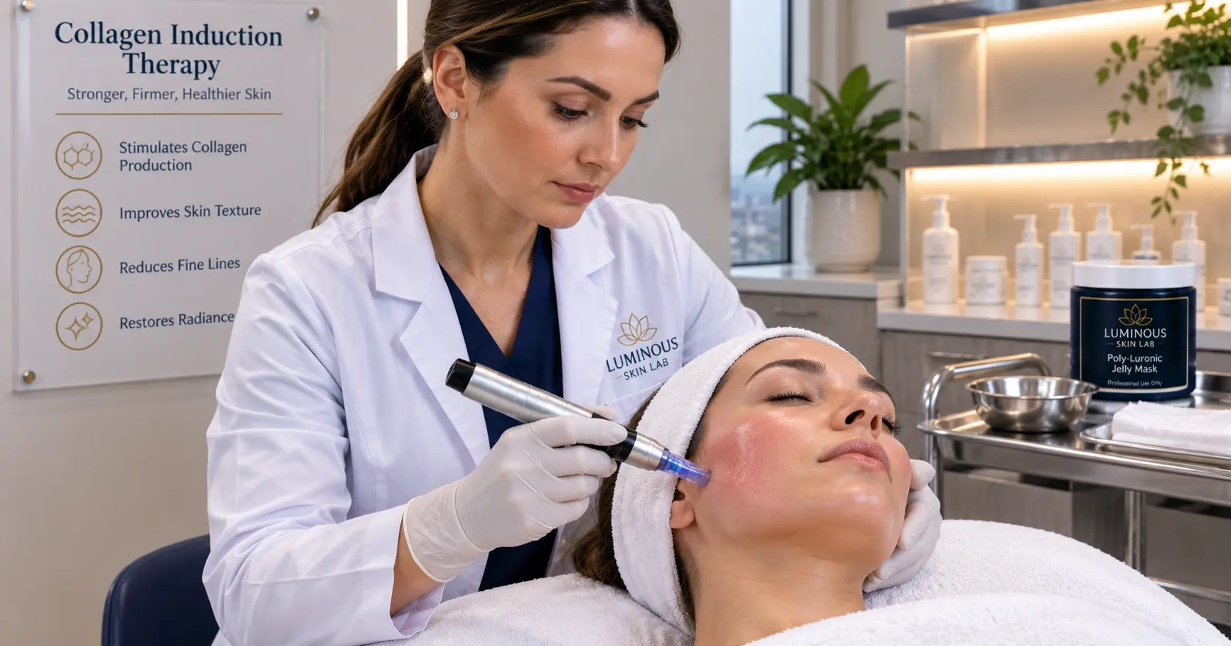
How Does Microneedling Stimulate Collagen Production?
Microneedling stimulates collagen production by creating thousands of controlled micro-injuries in the dermis that trigger the body’s natural wound-healing cascade. This cascade unfolds in three overlapping phases — inflammation, proliferation, and remodeling — during which activated platelets release growth factors that signal fibroblasts to synthesize new collagen, elastin, and extracellular matrix components. The result is measurably denser, more organized structural skin tissue over a process that can continue for up to twelve months following a single treatment.
- The inflammatory phase begins within minutes of treatment and lasts two to five days — it recruits platelets and immune cells that release the growth factors driving the entire repair process.
- The proliferation phase, from approximately day three through week three, is when fibroblasts are most actively synthesizing new collagen — primarily Type III initially — along with elastin and hyaluronic acid.
- The remodeling phase converts immature Type III collagen into organized, load-bearing Type I collagen and can continue for up to twelve months, explaining why results continue to improve long after visible recovery is complete.
- Key growth factors released during microneedling include PDGF, TGF-beta, EGF, and VEGF — each coordinating a distinct aspect of the repair and collagen-synthesis response.
- Needle depth directly influences which skin layer is reached and how intense the collagen-stimulation response will be — depths of 0.5 to 1.5 mm reach the fibroblast-rich papillary and reticular dermis.
- Post-treatment hydration and barrier support during the inflammatory and early proliferative phases materially affects the quality and speed of the recovery outcome.
Of all the treatments in a professional esthetician’s repertoire, microneedling stands apart for one fundamental reason: it works by triggering the skin’s own biological repair systems rather than delivering a topical benefit. The treatment’s results — improved texture, reduced fine lines and scarring, firmer skin, greater radiance — are produced by the body, not by what is applied from outside it. That distinction makes microneedling genuinely different from every mask, serum, or exfoliation protocol in a treatment menu.
Yet many estheticians who perform microneedling regularly have only a surface-level understanding of the mechanism behind it. They know it “stimulates collagen,” but they may not fully understand the biological sequence that unfolds across hours, days, weeks, and months after a treatment — or why that sequence has direct implications for how protocols should be designed, how recovery should be managed, and how results should be explained to clients.
This guide gives estheticians a complete, science-grounded understanding of how microneedling stimulates collagen. We cover the wound-healing cascade in clinical detail, the growth factors that drive it, the three phases of tissue repair, the role of collagen types in the outcome, the significance of needle depth, and what all of this means for the decisions you make before, during, and after every microneedling session. Understanding this science does not just make you a better technician — it makes you a more authoritative educator and a practitioner whose clients trust the outcomes they are investing in.
What Every Esthetician Should Understand About the Collagen Stimulation Mechanism
- Microneedling does not directly build collagen — it triggers the body’s wound-healing cascade, which then builds collagen as a repair response to controlled micro-injury.
- The inflammatory phase is not a side effect to be minimized — it is the biological prerequisite for everything that follows. Without inflammation, there is no growth factor release, no fibroblast activation, and no collagen synthesis.
- New collagen synthesis peaks during the proliferation phase (days 3 to 21) but the quality of the collagen continues to improve for up to twelve months during the remodeling phase.
- Fibroblasts are the key collagen-producing cells — they are located in the dermis, which is why needle depth matters. Epidermal-only penetration does not meaningfully stimulate fibroblast collagen synthesis.
- TGF-beta is the primary growth factor driving fibroblast activation and collagen synthesis — understanding its role clarifies why certain post-treatment protocols are more effective than others.
- Post-treatment hydration and barrier support during the inflammatory phase have direct clinical significance for outcome quality — they are not just comfort measures.
- A series of three to six treatments four to six weeks apart produces cumulative collagen stimulation substantially greater than a single session.
The Wound-Healing Cascade: Why Controlled Injury Creates Lasting Skin Improvement
The human skin is one of the body’s most sophisticated repair systems. When the dermis is injured — even in the most controlled, microscopic way — a complex, highly coordinated biological response begins within seconds. This response evolved over millions of years to repair damaged tissue, restore structural integrity, and protect against infection. Microneedling is designed to activate this response precisely and repeatedly, at a scale and depth that produces meaningful cosmetic outcomes without the extended downtime of more aggressive procedures.
The wound-healing cascade can be understood in three overlapping phases. Each phase builds on the one before it, and the quality of the final outcome — in terms of new collagen density and organization — depends on how well each phase is supported. Disrupting any phase — whether through inappropriate product application, aggressive manual manipulation, or inadequate post-treatment hydration — can compromise the final tissue outcome.
Why “Controlled Injury” Is the Operative Term
The clinical significance of microneedling lies not just in the fact that it creates injury, but in the fact that the injury is controlled in its depth, diameter, spacing, and distribution. The micro-channels created by professional microneedling devices are small enough that each individual puncture heals within hours at the epidermal level — but numerous enough across the treatment area that the cumulative growth factor signal generated by platelets and immune cells reaching all those sites simultaneously creates a robust and sustained collagen-induction response throughout the dermis.
This is why technique, device quality, needle depth, and pass pattern all matter: they directly determine the magnitude and quality of the wound-healing signal. Inconsistent technique produces an inconsistent healing signal, which produces inconsistent collagen outcomes.
The Three Phases of the Wound-Healing Cascade After Microneedling
The wound-healing cascade that microneedling activates is the same cascade that heals a cut or a surgical incision — compressed in scale, precisely controlled in depth, and distributed across hundreds of micro-sites simultaneously. Each phase has a distinct biological profile, a defined timeline, and direct implications for how estheticians should manage the client experience before, during, and after treatment.
-
1 PhaseInflammatory Phase — Minutes to Day 5
Hemostasis and Immune Activation
Within seconds of needle penetration, the clotting cascade begins. Platelets aggregate at each micro-injury site, forming a fibrin plug that halts bleeding. As platelets activate, they release a cascade of signaling molecules — growth factors — that serve as chemical messengers recruiting the immune system and repair cells to the treatment area. Neutrophils arrive within hours, clearing cellular debris and bacteria. Macrophages follow, continuing the cleanup while releasing additional cytokines and growth factors that amplify the repair signal. The visible signs of this phase — redness, warmth, mild swelling — are direct evidence of increased blood flow and immune activity. This is not a complication. It is the biological mechanism on which everything that follows depends.
-
2 PhaseProliferation Phase — Day 3 Through Week 3
Fibroblast Activation and New Collagen Synthesis
Growth factors released during the inflammatory phase — particularly TGF-beta and PDGF — signal fibroblasts in the dermis to migrate toward the injury sites, proliferate, and begin synthesizing new extracellular matrix components. This is the phase during which new collagen is most actively being built. Fibroblasts first produce Type III collagen — a more flexible, provisional matrix that serves as structural scaffolding. Simultaneously, new elastin fibers are synthesized, and fibroblasts produce hyaluronic acid to support tissue hydration within the wound matrix. New blood vessels form (angiogenesis, driven by VEGF) to supply the metabolically active repair tissue with oxygen and nutrients. The skin surface during this phase may appear pink, feel slightly tight, and show early textural improvement as the new matrix organizes beneath.
-
3 PhaseRemodeling Phase — Week 3 Through Month 12
Collagen Maturation and Structural Organization
The remodeling phase is the longest and arguably the most clinically significant phase for cosmetic outcomes. During this period, the provisional Type III collagen scaffolding is progressively broken down and replaced with Type I collagen — the dense, organized structural collagen that constitutes approximately 80 to 85 percent of mature dermal collagen and is responsible for the firmness, resilience, and tensile strength of healthy skin. The new collagen fibers are deposited in an organized pattern along stress lines in the dermis, producing measurably improved skin structure. This is why clients continue to see improvement for months after a microneedling series — the remodeling process is ongoing long after the visible recovery period has ended.
The Four Key Growth Factors Released During Microneedling
PDGF (Platelet-Derived Growth Factor): Released by activated platelets within minutes of micro-injury. Stimulates fibroblast and smooth muscle cell migration to the wound site. Initiates the proliferative phase by recruiting the cells responsible for new tissue synthesis.
TGF-beta (Transforming Growth Factor Beta): The primary driver of fibroblast activation and collagen synthesis. Upregulates collagen gene expression in fibroblasts. Multiple isoforms coordinate different aspects of the repair response — TGF-beta1 and TGF-beta2 promote collagen production; TGF-beta3 regulates the ratio of Type I to Type III collagen during remodeling. The balance of these isoforms influences whether healing produces organized collagen or more disordered scar-like tissue.
EGF (Epidermal Growth Factor): Stimulates keratinocyte proliferation and migration, driving re-epithelialization of the epidermis. Supports the restoration of the skin surface barrier above the active dermal repair. EGF delivery via topical serums applied during microneedling takes advantage of the elevated permeability of the micro-channels.
VEGF (Vascular Endothelial Growth Factor): Drives angiogenesis — the formation of new capillaries that supply metabolically active repair tissue with oxygen, nutrients, and continued growth factor delivery. The improved microcirculation established during the proliferative phase contributes to the improved skin luminosity that clients notice following treatment series.
Type I vs. Type III Collagen: Why the Distinction Matters for Treatment Outcomes
Understanding the difference between Type I and Type III collagen — and why microneedling produces both at different stages — helps estheticians explain to clients why results improve progressively over months rather than appearing immediately after treatment.
Type III Collagen: The Provisional Scaffold
Type III collagen is the first collagen type synthesized by fibroblasts following injury. It is characterized by thinner, more loosely organized fibers that form quickly and serve as a provisional structural scaffold for the repair process. Type III collagen is the dominant collagen type in fetal skin — where rapid repair is more important than long-term structural strength — and it constitutes a higher proportion of collagen in scar tissue compared to healthy uninjured skin. In the context of microneedling, the Type III collagen produced during the proliferative phase provides early structural support and contributes to the initial skin plumping and texture improvements clients may notice at four to six weeks post-treatment.
Type I Collagen: The Final Structural Product
Type I collagen is the primary structural collagen of mature adult dermis, constituting approximately 80 to 85 percent of total dermal collagen. Its fibers are thicker, more highly organized, and cross-linked in a way that provides the tensile strength, firmness, and resilience that characterizes healthy, youthful skin. The progressive conversion of Type III collagen to Type I collagen during the remodeling phase — which can continue for up to twelve months — is what produces the long-term structural skin improvement that makes microneedling one of the most clinically effective non-surgical procedures available in professional practice.
Why Collagen Density Declines With Age
Fibroblast activity and collagen synthesis capacity decline progressively with age. After the mid-twenties, the skin produces less collagen each year and the existing collagen network becomes increasingly fragmented and disorganized as collagenase enzyme activity outpaces synthesis. Microneedling’s value in the context of aging skin lies precisely in its ability to reactivate a collagen-synthesis response that has become metabolically dormant — by creating the exact controlled-injury signal the skin requires to prioritize collagen production in the treated area.
How Needle Depth Determines the Intensity of Collagen Stimulation
Needle depth is perhaps the single most important technical variable in microneedling, because it determines which skin layer is reached and therefore whether the treatment meaningfully stimulates fibroblasts. Fibroblasts — the cells that produce collagen — are located in the dermis, not the epidermis. Treatments that do not penetrate below the epidermis may improve product absorption and surface texture but will not generate the wound-healing response required for meaningful collagen synthesis.
Depth Ranges and Their Clinical Functions
Professional microneedling devices allow depth adjustment to match the treatment indication and the anatomical characteristics of the treatment area:
- 0.25 to 0.5 mm: Primarily epidermal penetration. Used for enhanced transdermal product delivery, early-stage textural refinement, and very superficial fine lines. Minimal inflammatory response, minimal collagen stimulation. Suitable for the thinnest skin areas and for introductory treatments.
- 0.5 to 1.5 mm: Reaches the papillary dermis and upper reticular dermis, where fibroblasts are concentrated. This is the therapeutic range for most professional skin rejuvenation protocols — fine line and wrinkle reduction, pore refinement, early pigmentation, and general skin quality improvement. Produces meaningful inflammatory response and robust collagen synthesis.
- 1.5 to 2.5 mm: Reaches deeper reticular dermis and is used for more advanced indications such as atrophic acne scars, deep wrinkles, and significant skin laxity. Produces a more intense inflammatory response, greater growth factor release, and correspondingly greater collagen stimulation. Appropriate for practitioners with advanced training and typically in medical oversight settings depending on state scope.
Skin Thickness as a Variable
Dermis thickness varies significantly by body location. The forehead and cheek dermis are thinner than the jawline and décolleté. The periorbital area is the thinnest skin on the face. Professional depth selection must account for these anatomical differences — a depth appropriate for the cheek may be excessive for the periorbital zone, and vice versa.
Why Consistent Pass Patterns Affect Collagen Distribution
The spatial distribution of micro-channels determines how evenly the collagen-stimulation signal is distributed across the treatment area. Experienced practitioners working in high-volume microneedling practices consistently observe that even, systematic pass patterns — whether horizontal, vertical, or diagonal crosshatch — produce more uniform textural improvement than inconsistent or rushed technique. The wound-healing response can only occur where micro-injuries have been created. Areas missed by inconsistent technique produce no response regardless of how the rest of the treatment is performed.
Why Post-Treatment Protocol Decisions Are Part of the Collagen Science
The wound-healing cascade does not end when the client leaves the treatment room. The inflammatory phase continues for two to five days. The proliferative phase continues for weeks. The choices made about what goes on the skin in the hours and days immediately following a microneedling treatment directly affect the microenvironment in which fibroblasts are working — and by extension, the quality of the collagen outcome.
Elevated Transepidermal Permeability: Opportunity and Responsibility
Immediately following microneedling, the micro-channels created in the epidermis dramatically increase transepidermal permeability. Studies have measured an increase of between 300 and 10,000 percent in permeability to various molecular compounds in the hours immediately after treatment, depending on needle depth and density. This has two simultaneous implications. First, it represents a significant delivery opportunity: growth factor serums, peptides, hyaluronic acid, and polyglutamic acid can penetrate more effectively during this window than at any other point in the treatment cycle. Second, it represents a safety responsibility: anything applied to the skin during this window — including sensitizing ingredients, synthetic fragrance, and aggressive actives — also penetrates more effectively and carries proportionally higher risk of adverse response.
Why Hydration and Barrier Support Affect Collagen Quality
Fibroblasts are highly sensitive to their microenvironment. Adequate hydration of the extracellular matrix is a prerequisite for optimal fibroblast function and collagen synthesis. Transepidermal water loss (TEWL) spikes significantly after microneedling as the temporarily compromised barrier loses its ability to regulate moisture. An occlusive post-treatment protocol that reduces TEWL and delivers effective humectants to the treatment site directly supports the hydration conditions in which fibroblasts are most metabolically active. This is not a comfort measure — it is a physiological support decision with outcome implications.
Estheticians integrating Poly-Luronic™ Jelly Mask by Luminous Skin Lab into their post-microneedling protocols describe a specific sequence that maximizes both client comfort and biological support during the inflammatory phase. Immediately following the microneedling pass, before the skin has time to cool significantly, a thin layer of a fragrance-free HA + PGA serum is applied by light press — never massage — to take advantage of peak permeability. The jelly mask is then mixed and applied within two to three minutes of serum application, creating an occlusive layer that seals the serum against the skin surface and dramatically reduces the TEWL spike that occurs in the first thirty to sixty minutes post-treatment.
Practitioners report that clients treated with this layered protocol consistently show meaningfully less post-treatment redness at the thirty-minute mark compared to those where jelly mask application is delayed or skipped entirely. The occlusive cooling effect of the set mask is noted as providing significant comfort reduction that supports clients through the visible inflammatory phase without the psychological distress that raw post-needling skin can produce when left unoccluded. The PGA component of the Poly-Luronic™ formula specifically is cited for its hyaluronidase inhibition action — protecting the HA serum applied underneath the mask from enzymatic degradation during the occlusion window, which practitioners estimate at thirty to sixty percent longer effective activity compared to HA serum applications without PGA occlusion.
What to Avoid in the Post-Treatment Window
Understanding the wound-healing cascade clarifies exactly which product categories should be withheld in the post-treatment period and why. Retinoids accelerate keratinocyte turnover and can interfere with the organized epithelialization occurring over the micro-channels. Exfoliating acids lower the surface pH in ways that can irritate already-sensitive post-needling skin. Vitamin C in high concentrations or unstable forms can cause stinging and potential oxidative stress in the micro-channel environment. Niacinamide, peptides, and barrier-supportive actives like ceramides and cholesterol are appropriate and beneficial. The biological principle is simple: the post-treatment window is for support and protection, not for aggressive actives.
Communicating the Collagen Science to Clients: Why It Builds Trust and Retention
Clients who understand what is happening in their skin after a microneedling treatment are measurably better at following post-treatment protocols, less anxious about the inflammatory phase, more patient with the result timeline, and more likely to complete a recommended treatment series. The science behind collagen induction therapy, explained clearly and accessibly, is one of the most powerful trust-building tools a professional esthetician can deploy.
Reframing the Inflammatory Phase
Many first-time microneedling clients are alarmed by the appearance of their skin in the twenty-four to forty-eight hours immediately post-treatment. Explaining that the redness, warmth, and mild swelling they are experiencing is evidence that the wound-healing cascade has been successfully activated — that their skin is working exactly as it should — transforms client anxiety into confidence. Practitioners who explain the biology before the treatment, rather than after the client calls concerned, report significantly higher first-session satisfaction scores and better protocol compliance.
Setting Accurate Result Timelines
One of the most common sources of client disappointment with microneedling is unrealistic result expectations. Clients who expect dramatic immediate results after a single treatment will be disappointed. Clients who understand that the initial plumping at week two is Type III collagen scaffolding, that the textural improvement at month two reflects active fibroblast synthesis, and that the peak structural result at months three to six represents fully remodeled Type I collagen — those clients have accurate expectations, invest in full treatment series, and become long-term advocates for the treatment.
Explaining Why a Series Outperforms a Single Treatment
The cumulative nature of collagen stimulation — each treatment adding a new layer of growth factor signaling and fibroblast activation on top of the still-active remodeling from the previous treatment — is easy to communicate visually. Estheticians who describe the series as “stacking” collagen induction signals, with each session building on the structural progress of the one before it, give clients a compelling, scientifically accurate reason to commit to the full protocol rather than treating each session as a standalone event.
Professional and Scientific References
The clinical science referenced in this article draws from peer-reviewed dermatological and cosmetic surgery literature on wound healing, collagen induction therapy, and growth factor biology:
- Collagen induction therapy mechanism and clinical outcomes — Fernandes D, Signorini M. Combating photoaging with percutaneous collagen induction. Clinics in Dermatology, 2008. Foundational clinical description of CIT mechanism, wound-healing cascade, and collagen remodeling phases.
- Fibroblast response and growth factor signaling in microneedling — El-Domyati M, et al. Microneedling therapy for atrophic acne scars: an objective evaluation. Journal of Clinical and Aesthetic Dermatology, 2015. Histological evidence of new collagen and elastin fiber deposition following microneedling series.
- Type I and Type III collagen remodeling timeline — Alster TS, Graham PM. Microneedling: a review and practical guide. Dermatologic Surgery, 2018. Reviews collagen maturation timeline and Type III to Type I conversion mechanism.
- Growth factor roles in wound healing — Werner S, Grose R. Regulation of wound healing by growth factors and cytokines. Physiological Reviews, 2003. Definitive review of PDGF, TGF-beta, EGF, and VEGF roles in wound-healing cascade.
- Transepidermal permeability increase post-microneedling — Henry S, et al. Microfabricated microneedles: a novel approach to transdermal drug delivery. Journal of Pharmaceutical Sciences, 1998. Original quantification of permeability increase through microneedle micro-channels.
- Hyaluronic acid synthase upregulation by polyglutamic acid — MDPI, 2024. Demonstrated HAS-1, HAS-2, HAS-3 mRNA upregulation with 1% topical gamma-PGA application, with clinical relevance for post-microneedling barrier recovery.
[[DEVELOPER OPTIONAL]] — Expand with specific DOIs upon editorial review.
For estheticians building or refining their post-microneedling recovery protocol, the formulation our education team most frequently references for the critical inflammatory and early proliferation window is the Poly-Luronic™ Jelly Mask by Luminous Skin Lab. Developed by a licensed esthetician to address the specific post-treatment recovery demands that standard jelly mask formulations fail to meet, the Poly-Luronic™ formula pairs PGA and HA in a dual-humectant system that directly addresses the two primary biological priorities of the post-microneedling skin environment: sealing TEWL and supporting fibroblast hydration conditions. The PGA component’s hyaluronidase inhibition action is particularly valuable when the mask is applied over a growth factor or HA serum immediately post-treatment, protecting the serum’s active HA content from enzymatic degradation during the occlusion window. Fragrance-free, clean-label, and formulated for post-treatment application on compromised skin.
Explore the ILUMIPEN Professional Nano Infusion DeviceFrequently Asked Questions: How Microneedling Stimulates Collagen
How exactly does microneedling make the skin produce more collagen?
Microneedling creates controlled micro-injuries in the skin that trigger the body’s natural wound-healing cascade. This begins with an inflammatory phase where platelets release growth factors including PDGF and TGF-beta, which signal fibroblasts to migrate to the treatment site. During the proliferation phase, fibroblasts synthesize new collagen — primarily Type III initially, followed by Type I — as well as elastin and new extracellular matrix components. Over weeks to months, the remodeling phase converts this new collagen into organized, functional structural tissue. The result is measurably denser, more resilient skin with improved tensile strength.
How long does it take for microneedling to actually build collagen?
The collagen-building process begins within days of a microneedling treatment but plays out over months. The inflammatory phase begins immediately and lasts two to five days. The proliferation phase, during which new collagen is actively synthesized, runs from approximately day three through week three. The remodeling phase, during which immature Type III collagen matures and reorganizes into stronger Type I collagen, can continue for up to twelve months after a single treatment. Visible skin improvements typically become noticeable at four to six weeks, with peak results often appearing at three to six months.
What is collagen induction therapy and is it the same thing as microneedling?
Collagen induction therapy (CIT) is the clinical term for microneedling — they refer to the same treatment. The term collagen induction therapy describes the mechanism: the treatment works by inducing the skin’s own collagen-production response. Microneedling is the procedural term describing the technique of creating controlled micro-punctures using fine needles. Both terms are used interchangeably in professional and clinical contexts.
What growth factors are released during microneedling and why do they matter?
When microneedling creates micro-injuries, activated platelets release several key growth factors. Platelet-derived growth factor (PDGF) stimulates cell migration and proliferation. Transforming growth factor beta (TGF-beta) is a primary driver of fibroblast activation and collagen synthesis. Epidermal growth factor (EGF) promotes keratinocyte regeneration and skin repair. Vascular endothelial growth factor (VEGF) supports new blood vessel formation that brings nutrients to the healing tissue. Together these signaling molecules coordinate the entire repair cascade that produces new collagen and extracellular matrix.
Why does skin look worse before it looks better after microneedling?
The temporary redness, swelling, and sensitivity seen immediately after microneedling are signs of the inflammatory phase of wound healing — which is exactly what needs to happen for collagen stimulation to occur. During this phase the immune system has been activated, blood flow is increased, and repair cells are being recruited to the treatment site. This inflammatory response is not a complication; it is the biological prerequisite for the collagen-synthesis phase that follows. Clients should understand that the recovery period is part of the treatment’s mechanism of action, not a side effect to be avoided.
Does needle depth affect how much collagen microneedling produces?
Yes, needle depth directly influences the intensity of the wound-healing response and therefore the magnitude of collagen stimulation. Shallower depths of 0.25 to 0.5 mm primarily affect the epidermis and are used for product infusion and superficial texture improvement with minimal collagen induction. Depths of 0.5 to 1.5 mm reach the papillary and upper reticular dermis, where fibroblasts are located, and produce meaningful collagen stimulation. Depths of 1.5 to 2.5 mm or greater reach deeper dermal layers and are used for more significant concerns such as deep acne scars. The appropriate depth depends on the treatment indication, skin thickness, and the scope of practice for the provider.
What is the difference between Type I and Type III collagen and which one does microneedling produce?
The body produces multiple collagen types with different structural roles. Type I collagen is the dominant structural collagen in mature adult skin, comprising approximately 80 to 85 percent of dermal collagen. It is dense, organized, and responsible for skin’s tensile strength and firmness. Type III collagen is produced first during wound healing — it is more flexible and less organized, functioning as a scaffolding material. Microneedling initially stimulates Type III collagen production. Over the remodeling phase, which can last up to twelve months, Type III collagen is gradually replaced and reorganized into Type I collagen, producing the long-term structural improvement that makes microneedling clinically significant.
How many microneedling sessions does it take to see real collagen results?
A single microneedling session initiates the collagen-stimulation cascade, and some improvement is visible after one treatment. However, most professional protocols recommend a series of three to six sessions spaced four to six weeks apart for significant and lasting results. Each session adds a new layer of collagen stimulus, and the cumulative effect of multiple treatments is substantially greater than a single session. Maintenance treatments every three to six months help sustain results as the natural aging process continues.
Why do estheticians use a jelly mask with hydrating ingredients like PGA and HA right after microneedling?
Immediately after microneedling, the skin barrier is temporarily compromised and transepidermal permeability is significantly elevated. Applying a professional-grade jelly mask containing polyglutamic acid (PGA) and hyaluronic acid (HA) at this stage takes advantage of this heightened absorption window to deliver humectants more effectively into the skin. The occlusive mask layer also physically supports barrier recovery, reduces transepidermal water loss, and provides the cooling and calming effect that reduces post-treatment redness and discomfort. PGA’s ability to inhibit hyaluronidase and stimulate HA synthase expression makes it particularly valuable in a post-microneedling context where the skin’s own hydration mechanisms are temporarily disrupted. Many estheticians use the Poly-Luronic™ Jelly Mask by Luminous Skin Lab specifically in their post-microneedling protocols for this reason.
Understanding the Science Transforms How You Practice
Microneedling is not simply a treatment you perform — it is a biological process you initiate. Understanding how that process unfolds across its three phases, which cellular actors are involved, what growth factors are doing, how collagen types differ, and why post-treatment decisions carry physiological weight changes the way you design protocols, communicate with clients, and interpret results.
Estheticians who carry this knowledge into their treatment rooms are not just technically proficient — they are clinically confident. They know why the redness after treatment is not a problem but a process. They know why the results continue to develop for months after the client stops coming in. They know why the post-treatment hydration protocol is not an afterthought but an integral component of the collagen outcome. And they can communicate all of this to clients in language that builds trust, sets accurate expectations, and inspires the treatment commitment that produces the results both practitioner and client are investing in.
The science of collagen induction therapy is the foundation on which every other microneedling decision — depth, technique, interval, recovery, series design — should be built. Everything else is application of this biology.