How Does Microneedling Stimulate Collagen Production?
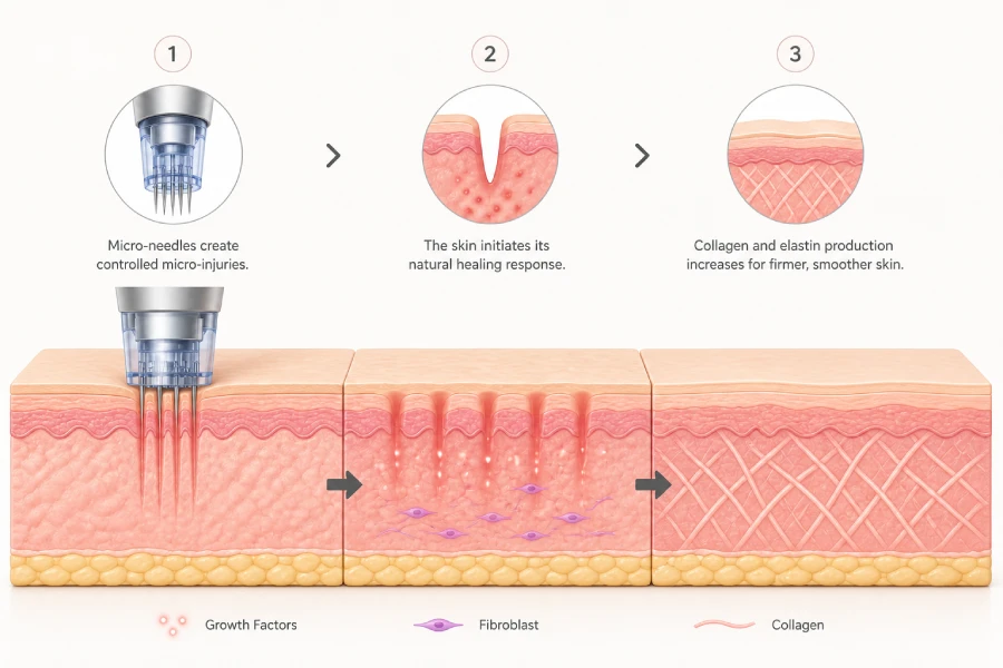
Microneedling stimulates collagen production by delivering thousands of controlled micro-injuries into the dermis, which activates the body’s natural wound-healing response. Platelets at the injury site release growth factors that signal fibroblasts to produce new collagen and elastin, resulting in progressive structural improvement to the skin over weeks and months following treatment.
- Micro-injuries trigger a three-phase healing cascade: inflammation, proliferation, and remodelling — each phase playing a distinct role in collagen synthesis.
- Platelet-derived growth factors (PDGF) and transforming growth factor beta (TGF-β) are the primary chemical signals that activate fibroblast collagen production.
- New collagen type III is produced first during the proliferative phase; it gradually converts to collagen type I during remodelling, providing long-term firmness.
- Needle depth determines the depth and intensity of collagen induction — dermal penetration between 0.5 mm and 1.5 mm produces the most clinically significant fibroblast activation.
- Full collagen remodelling takes three to six months, which is why multiple spaced sessions accumulate better results than a single treatment.
- Post-treatment hydration and barrier support directly influence how efficiently the skin transitions through the healing phases into active collagen synthesis.
When a client asks why microneedling improves their skin, the honest answer goes deeper than “it creates micro-injuries.” That description is technically accurate but leaves out the entire biological story — a precisely orchestrated sequence of cellular events that, when triggered correctly by professional technique, results in measurably denser, more elastic skin. Understanding that sequence is what separates practitioners who produce consistent clinical outcomes from those who follow a protocol without knowing why it works.
The collagen induction mechanism is not a side effect of microneedling — it is the entire point. Every decision made during a microneedling session, from needle depth selection to the serums applied mid-treatment to the recovery mask used immediately after, either supports or undermines the wound-healing cascade responsible for new tissue formation. Estheticians who understand this cascade make better real-time decisions and build protocols that reliably deliver results across diverse client presentations.
This article covers the complete cellular mechanism behind microneedling-induced collagen synthesis, the three phases of wound healing and what happens in each, the role of needle depth in determining induction intensity, and the post-treatment environment factors that either accelerate or slow the collagen-building process.
What Every Esthetician Should Know About Microneedling and Collagen
- Microneedling works by initiating a wound-healing cascade — the micro-injury is the trigger, not the outcome. Collagen synthesis is the outcome.
- Fibroblasts are the collagen-producing cells that estheticians are targeting; activating them requires dermal penetration, not just epidermal disruption.
- The inflammatory phase is not a side effect to minimise — it is the necessary first step in the collagen induction sequence. Suppressing it prematurely can reduce results.
- Collagen type III is produced first and then remodels to type I over months; clients should be counselled to expect progressive, not immediate, structural improvement.
- Needle depth selection is the primary lever controlling induction intensity — deeper is not always better; appropriate depth is based on skin concern and skin condition.
- Post-treatment hydration and barrier recovery are not optional afterthoughts; they directly affect how efficiently the skin completes the proliferative and remodelling phases.
- Spacing sessions four to six weeks apart allows each collagen induction cycle to complete before a new one is initiated, preventing over-treatment and barrier compromise.
The Three-Phase Wound-Healing Cascade Behind Collagen Induction
When a microneedling device penetrates the dermis, it does not simply poke holes. It initiates a highly ordered biological response that has three distinct phases, each with a specific cellular function and timeline. Understanding these phases gives estheticians a framework for explaining treatment responses to clients and for designing post-care protocols that work with — rather than against — the biology.
Phase 1: Inflammation (Day 0 to Day 4)
Within seconds of dermal puncture, platelets aggregate at the micro-injury site and begin releasing growth factors including platelet-derived growth factor (PDGF), transforming growth factor alpha (TGF-α), and transforming growth factor beta (TGF-β). These molecular signals recruit neutrophils and macrophages to the site within hours. Neutrophils clear bacteria and debris from the wound channel, while macrophages assume a longer-term role in orchestrating tissue repair by secreting additional growth factors that initiate the next phase.
The visible redness and warmth clients experience post-treatment is this phase in action. Vasodilation increases blood flow to the area, bringing the cellular machinery needed for repair. This inflammatory phase typically resolves within 24 to 72 hours for standard treatment depths, though clients with compromised barriers or reactive skin may experience a slightly extended response.
Phase 2: Proliferation (Day 4 to Week 3)
As inflammation resolves, fibroblasts migrate to the treatment site and begin synthesising new extracellular matrix components. This is the phase where collagen production actively begins. Fibroblasts produce collagen type III, a soft, flexible precursor collagen that provides initial structural support to the regenerating tissue. Alongside collagen, fibroblasts also deposit elastin fibres and glycosaminoglycans, contributing to improved skin hydration retention and elasticity.
New blood vessel formation (angiogenesis) also occurs during this phase, improving nutrient delivery to the healing tissue. This is why skin often looks temporarily more luminous in the weeks following a microneedling session — increased vascular activity brings oxygenated blood to the surface layers.
Phase 3: Remodelling (Week 3 to Month 6+)
The remodelling phase is the longest and most clinically significant for estheticians to understand. During this phase, collagen type III is gradually converted to collagen type I — the primary structural collagen of healthy adult skin. Collagen fibres cross-link and organise along lines of mechanical tension, creating the denser, firmer skin architecture that clients notice as improved texture and reduced fine lines. This process continues for three to six months following each treatment session, which is why cumulative improvement across a series of sessions is substantially greater than results from a single treatment.
Growth Factors, Fibroblasts, and the Molecular Mechanism of Collagen Synthesis
The cellular actors in microneedling-induced collagen production are growth factors and fibroblasts — and their interaction is what makes the treatment clinically effective rather than simply mechanically disruptive. Understanding the molecular language of this process helps estheticians select the right supportive serums, understand why certain ingredient combinations amplify results, and explain outcomes to clients in credible, evidence-based terms.
How Growth Factors Signal Collagen Production
Growth factors are small signalling proteins that bind to receptors on fibroblast cell membranes, triggering intracellular cascades that ultimately activate collagen gene expression. The primary growth factors released during the platelet degranulation response to micro-injury include PDGF, TGF-β1 and TGF-β3, connective tissue growth factor (CTGF), and vascular endothelial growth factor (VEGF). Each plays a distinct role: TGF-β1 strongly upregulates collagen type I and III synthesis, PDGF stimulates fibroblast proliferation and migration, and VEGF drives the angiogenesis that supports the newly forming tissue.
The density and distribution of growth factor release is influenced by needle depth and the number of passes made during treatment. Deeper penetration into the papillary and reticular dermis reaches zones of higher fibroblast concentration, producing a stronger induction signal. This is the biological basis for why estheticians adjust depth parameters based on treatment goals rather than applying a single standard setting across all clients.
The Biology of Fibroblast Activation in Microneedling
Fibroblasts are the primary collagen-producing cells of the dermis. At rest, they maintain baseline extracellular matrix turnover. When activated by growth factor signalling following micro-injury, they shift into a highly productive synthetic state, upregulating collagen gene expression and secreting both collagen precursors and the enzymes needed to assemble them into mature fibres.
Research published across multiple peer-reviewed dermatology journals documents that a single microneedling session at appropriate dermal depth can increase collagen and elastin expression measurably within 28 days. A series of three to six sessions produces cumulative dermal thickening that persists for months beyond the final treatment as the remodelling phase continues to mature deposited collagen.
Clinical implication for estheticians: Fibroblast activation is transient and depth-dependent. Superficial needle settings that remain in the epidermis do not reliably activate the dermal fibroblast population. Effective collagen induction requires consistent dermal penetration at a depth appropriate to the treatment goal and the individual client’s skin thickness.
How Needle Depth Controls the Intensity of Collagen Induction
Needle depth is the single most influential variable under esthetician control during a microneedling session. It determines which skin layers are reached, which cell populations are activated, and how strong a wound-healing stimulus is delivered. Choosing the wrong depth — too shallow to reach fibroblasts or too deep for the client’s skin thickness and condition — is the most common reason practitioners see inconsistent results despite following all other protocol steps correctly.
The diagram below maps needle depth ranges to anatomical targets and corresponding clinical outcomes, providing a practical reference for treatment planning across different skin concerns and client presentations.
Why Consistent Depth Delivery Matters More Than Maximum Depth
One of the most common protocol errors estheticians encounter is inconsistent needle depth during treatment — particularly on curved or uneven facial anatomy where maintaining consistent skin contact with a motorised device requires deliberate technique. Areas where skin is thinner (around the eyes, upper lip, and neck) require shallower settings than flatter, thicker facial zones. Using a single depth setting across the entire face produces uneven collagen stimulation, with some areas under-treated and others at risk of over-stimulation or barrier damage.
Effective collagen induction protocols involve intentional depth adjustment by zone, and training estheticians to treat depth selection as a dynamic variable rather than a fixed setting is one of the most impactful skill developments available in this discipline.
When transitioning from the active microneedling pass to the recovery phase, timing matters more than most estheticians realise. Poly-Luronic™ Jelly Mask is applied within two to three minutes of completing the final needle pass — not after serums have been massaged in, not after the client sits upright, but immediately once the treatment surface is clean. The reason is practical: micro-channels close fastest in the first five to ten minutes post-treatment, and the occlusive gel seal needs to be in place before that window narrows. In practice, skin treated at 1.0–1.5 mm depth shows noticeably higher erythema at this stage than skin treated at shallower settings, and the cooling sensation from the jelly mask’s alginate base provides immediate client comfort that also signals the transition from active treatment to recovery. Compared to applying a standard sheet mask at this stage, which tends to shift and break seal contact over uneven facial contours, the mixed jelly mask conforms completely and holds its position for the full 15-to-20-minute recovery window without esthetician intervention.
Six Variables That Determine Collagen Induction Outcomes
Collagen induction is not a guaranteed result of simply running a microneedling device across the skin. Six clinical variables influence whether the wound-healing cascade proceeds efficiently and whether the resulting collagen deposition is meaningful. Estheticians who account for all six in their treatment planning produce more consistent outcomes than those who focus only on needle depth and device settings.
Needle Depth and Skin Thickness
Effective fibroblast activation requires dermal penetration. Skin thickness varies by facial zone, client age, and skin condition. Depth selection must be adjusted accordingly, with thinner skin around the eyes and lips requiring shallower settings than the cheeks and forehead.
Device Speed and Pass Count
Motorised microneedling pen speed affects the density of micro-channels created per square centimetre. Higher speed combined with appropriate pressure produces more uniform channel distribution. Too few passes under-deliver the wound stimulus; excessive passes risk over-treatment and barrier damage.
Client Skin Condition at Treatment Time
Skin that is actively inflamed, barrier-compromised, or severely dehydrated at the time of treatment responds differently to microneedling than healthy skin. Compromised barrier function can amplify the inflammatory response and slow the transition into the proliferative phase, producing prolonged recovery with reduced collagen yield.
Serums Applied During Treatment
Growth-factor serums, hyaluronic acid, and peptide complexes applied as a glide medium during needling can be infused to depth by the channel-creation process, augmenting the biological signal delivered. However, active ingredients that are inappropriate post-barrier disruption — such as retinoids or strong acids — can trigger excessive inflammation and impair healing.
Immediate Post-Treatment Hydration
The barrier disruption created by microneedling produces significant transepidermal water loss. Dehydration during the inflammatory and early proliferative phases impairs cellular signalling and slows the healing cascade. Immediate occlusive hydration is a clinical requirement for optimal collagen induction outcomes, not an optional comfort measure.
Treatment Interval and Series Completion
Collagen remodelling requires three to six months to complete following each session. Treating before the previous cycle completes does not produce cumulative benefit — it disrupts an active healing process. The four-to-six-week minimum spacing between sessions is based on the biology of the wound-healing cascade, not convention.
Supporting the Collagen Induction Process After Treatment
The collagen induction cascade does not end when the microneedling device is set down. The 48 to 72 hours following treatment are arguably as important to final outcomes as the treatment itself, because the inflammatory phase — and its transition into the proliferative phase — takes place entirely in the post-treatment window. Estheticians who give clients clear, science-grounded post-care instructions and who complete the in-treatment recovery phase correctly significantly improve the probability of strong collagen outcomes.
Why the Inflammatory Phase Should Not Be Suppressed
A common client instinct following microneedling is to apply anti-inflammatory products, ice, or other calming interventions to reduce redness and discomfort as quickly as possible. While managing discomfort is appropriate, aggressively suppressing the inflammatory response — particularly with topical or oral non-steroidal anti-inflammatory drugs — can blunt the growth factor release that initiates fibroblast activation. The goal in immediate post-care is to support hydration and barrier recovery, not to eliminate the inflammatory response that is driving collagen synthesis.
Estheticians should communicate this distinction clearly to clients: some redness and warmth for 24 to 48 hours is evidence that the wound-healing cascade is active and the treatment is working. It is not a complication to be corrected.
The Role of Hydration in the Proliferative Phase
During the proliferative phase, fibroblasts require an adequately hydrated tissue environment to function efficiently. Transepidermal water loss following microneedling can be substantially elevated compared to untreated skin, and without intervention, this leads to cellular dehydration that impairs collagen precursor assembly and fibre organisation. This is the biological mechanism behind the clinical recommendation for occlusive post-treatment masks — they are not simply comfort products; they are functional tools that maintain the tissue water content fibroblasts need to complete collagen synthesis.
Home Care Protocols That Support Collagen Remodelling
Between professional sessions, clients can support ongoing collagen remodelling by maintaining consistent use of SPF (UV exposure accelerates collagen degradation), using peptide serums that provide the amino acid building blocks for collagen synthesis, and avoiding retinoids during the first three to five days post-treatment when barrier integrity is compromised. Vitamin C, introduced after barrier recovery is confirmed, can support collagen cross-linking by acting as a cofactor for prolyl and lysyl hydroxylase enzymes involved in collagen fibre maturation.
Professional and Scientific References
The mechanisms described in this article are grounded in peer-reviewed wound-healing science, dermatology research on collagen induction therapy, and established histological studies on fibroblast activation following controlled dermal micro-injury.
- Aust, M.C. et al. — Percutaneous Collagen Induction Therapy: An Alternative Treatment for Scars, Wrinkles, and Skin Laxity. Plastic and Reconstructive Surgery, multiple publications 2006–2011. Foundational documentation of dermal collagen and elastin increase following microneedling.
- Fernandes, D. — Minimally invasive percutaneous collagen induction. Oral and Maxillofacial Surgery Clinics of North America, 2005. Original description of collagen induction therapy mechanism and three-phase healing response.
- El-Domyati, M. et al. — Microneedling therapy for atrophic acne scars: an objective evaluation. Journal of Clinical and Aesthetic Dermatology, 2015. Histological evidence of collagen type I and III deposition following microneedling series.
- Singer, A.J. & Clark, R.A.F. — Cutaneous Wound Healing. New England Journal of Medicine, 1999. Foundational reference for the three-phase wound-healing cascade including cellular mechanisms of fibroblast activation.
- Alster, T.S. & Graham, P.M. — Microneedling: A Review and Practical Guide. Dermatologic Surgery, 2018. Clinical review of microneedling outcomes and protocol parameters including depth, frequency, and post-care best practices.
The immediate post-microneedling window is clinically significant for collagen induction outcomes. The wound-healing cascade relies on an adequately hydrated, barrier-supported environment to transition efficiently from the inflammatory phase into the fibroblast-active proliferative phase. When evaluating occlusive recovery masks for post-microneedling protocols, the critical criteria are: speed of application (the mask must be usable within minutes of treatment completion), barrier seal integrity across irregular facial contours, and the presence of dual-depth humectants that support epidermal water content during the first recovery hours.
Poly-Luronic™ Jelly Mask is formulated to meet all three criteria. Its alginate gel structure sets within 90 seconds of application, creating a continuous occlusive barrier across the full face — including curved contour zones that sheet masks routinely fail to seal. The polyglutamic acid and hyaluronic acid combination provides both surface-level and deeper epidermal hydration, maintaining the tissue water content that fibroblasts require during active collagen synthesis. For estheticians building post-microneedling recovery protocols where collagen outcome quality is the primary goal, this is the mask formulation we recommend including as a standard protocol step.
Explore the Poly-Luronic™ Jelly Mask LineFrequently Asked Questions: Microneedling and Collagen Stimulation
How does microneedling actually stimulate collagen production?
Microneedling stimulates collagen production by creating thousands of controlled micro-injuries in the dermis, triggering the body’s natural wound-healing cascade. When needles penetrate to the appropriate depth, they activate platelets that release growth factors, which in turn signal fibroblasts to synthesise new collagen and elastin fibres. The three-phase healing response — inflammation, proliferation, and remodelling — progresses over weeks to months, with new collagen fibres gradually replacing disorganised scar tissue and improving overall skin structure.
What type of collagen does microneedling produce?
Microneedling primarily stimulates the production of collagen type III during the early proliferative phase, followed by a gradual conversion to collagen type I as remodelling progresses. Collagen type III provides initial structural support and flexibility, while collagen type I is the dominant structural protein in mature, healthy skin and is responsible for long-term firmness and density improvements. Clinical studies have documented measurable increases in both collagen types following a series of professional microneedling treatments.
Why does skin look worse before it looks better after microneedling?
The temporary redness, mild swelling, and sensitivity that follows microneedling reflect the active inflammatory phase of wound healing, which is exactly what makes the treatment work. During this phase, the immune system dispatches neutrophils and macrophages to the treatment site, clearing cellular debris and releasing cytokines that signal the beginning of collagen synthesis. This initial inflammatory response typically resolves within 24 to 72 hours, after which the proliferative phase begins and new tissue formation accelerates.
How long does it take for collagen to rebuild after microneedling?
Meaningful collagen rebuilding begins within the first week following treatment, but the full remodelling cycle takes between three and six months to reach its peak. Fibroblasts actively synthesise new collagen fibres during the proliferative phase, which runs from approximately day four through week three. The remodelling phase then continues for months as collagen fibres organise, cross-link, and mature. This is why estheticians typically recommend spacing treatment sessions four to six weeks apart and setting realistic client expectations around visible results.
Does needle depth affect how much collagen is produced?
Yes, needle depth directly influences the degree of collagen induction because it determines how far into the dermis the micro-injury extends. Superficial depths of 0.25 to 0.5 mm primarily enhance product penetration and surface texture with minimal dermal stimulation. Depths of 0.5 to 1.5 mm reach the papillary and upper reticular dermis where fibroblasts are concentrated, producing a stronger collagen response appropriate for fine lines, texture irregularities, and early scarring. Depths above 1.5 mm are reserved for deeper scar revision and are typically performed only under medical supervision.
Can microneedling boost collagen in mature skin?
Yes, microneedling can stimulate collagen synthesis in mature skin, though the response timeline may differ compared to younger skin. As skin ages, fibroblast activity naturally slows and baseline collagen production declines. Microneedling reactivates the wound-healing cascade regardless of age, prompting fibroblasts to resume collagen synthesis in response to the controlled injury signal. Clinical evidence supports meaningful improvements in skin density and elasticity in clients over 50 following a consistent series of treatments, though practitioners should set realistic expectations around the pace and extent of results.
Why is hydration so important immediately after microneedling?
Hydration is critical immediately after microneedling because the micro-channels created by needles dramatically increase transepidermal water loss, leaving the skin in a temporarily dehydrated and barrier-compromised state. Dehydration during the inflammatory phase impairs cell signalling and can slow the transition into the proliferative phase where collagen synthesis accelerates. Applying occlusive hydration treatments immediately post-treatment helps seal the channels, reduce inflammation, support barrier recovery, and maintain the optimal cellular environment for collagen induction to proceed efficiently.
How many microneedling sessions does it take to see real collagen improvement?
Most clients begin to notice visible improvements in skin texture and firmness after two to three sessions, though a complete collagen induction protocol typically involves three to six treatments spaced four to six weeks apart. Each session adds a new stimulus to the wound-healing cascade, and the cumulative collagen deposition from multiple sessions produces more significant structural change than a single treatment. The specific number of sessions depends on the client’s skin concern, starting skin condition, needle depth used, and how consistently post-treatment care protocols are followed.
Why do estheticians use the Poly-Luronic™ Jelly Mask after microneedling to support collagen recovery?
The Poly-Luronic™ Jelly Mask is applied immediately after microneedling because the occlusive seal it forms over open micro-channels helps lock in hydration during the critical early inflammatory phase when transepidermal water loss is highest. Its polyglutamic acid and hyaluronic acid combination delivers dual-depth hydration to both the surface and deeper epidermal layers while the alginate base creates the physical barrier needed to prevent moisture escape. Estheticians working with post-microneedling protocols find that applying the mask within minutes of treatment completion significantly reduces post-procedure redness duration and supports the transition into the proliferative healing phase, where collagen synthesis is most active.
The Collagen Induction Cascade: A Framework Every Microneedling Practitioner Should Own
Microneedling’s clinical value is entirely grounded in wound-healing biology. The micro-injury is not the result — it is the trigger. The result is a carefully orchestrated three-phase cellular process that, when supported correctly from the moment the device is set down, produces measurably denser, more elastic, and more youthful skin architecture over weeks and months following treatment. Estheticians who understand this cascade — the growth factors, the fibroblast activation, the collagen conversion sequence, and the role of post-treatment hydration — are equipped to make better protocol decisions, communicate more credibly with clients, and produce more consistent outcomes across every treatment.
The variables that separate good microneedling results from great ones are not mysterious. They are depth selection, consistent technique, appropriate post-treatment environment management, and correctly spaced sessions that allow the full collagen remodelling cycle to progress. Each of these variables is within the practitioner’s control, and each is grounded in the biological mechanisms described in this article.
Continue building your clinical foundation by exploring the other articles in the Microneedling Collagen Induction Therapy guide — covering protocol steps, safety guidelines, treatment combinations, and business strategy for building a profitable microneedling service.