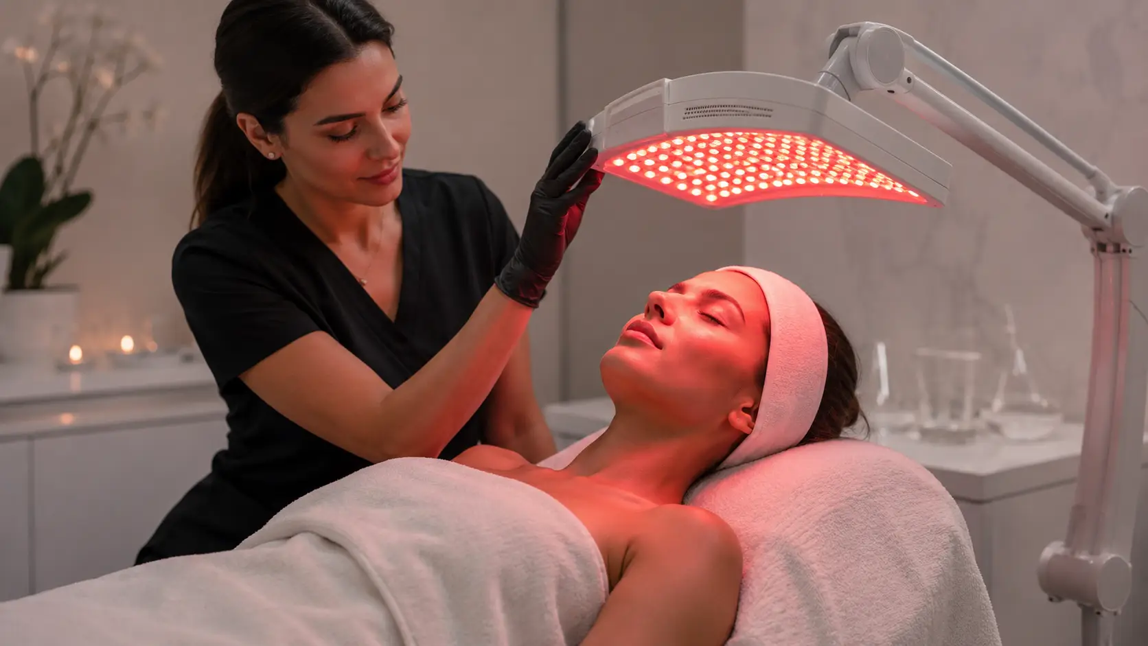
Does LED Light Therapy Stimulate Collagen Production?
Yes. Peer-reviewed research consistently confirms that red and near-infrared LED light therapy stimulates collagen production through a measurable, non-thermal cellular mechanism called photobiomodulation. Specific wavelengths of light are absorbed by mitochondria in skin fibroblasts, triggering a cascade of biochemical events that upregulate collagen type I and type III synthesis and suppress the enzymes responsible for collagen breakdown.
- Photobiomodulation works through cytochrome c oxidase in the mitochondria — when red or near-infrared photons are absorbed, cells produce more ATP, reduce oxidative stress, and activate downstream collagen synthesis pathways.
- The most clinically supported wavelengths for collagen stimulation are red light at 630–670nm and near-infrared at 830–850nm. Red light activates fibroblasts in the papillary dermis; near-infrared reaches deeper into the reticular dermis.
- Clinical studies document measurable increases in dermal collagen density following a structured series of treatments, typically 8 to 12 sessions delivered 2 to 3 times per week.
- LED therapy also suppresses matrix metalloproteinases (MMPs) — the enzymes that degrade existing collagen — providing a dual mechanism of both building new collagen and protecting existing structure.
- The collagen response is dose-dependent: irradiance level, energy dose, wavelength, and treatment frequency all directly determine clinical outcomes. Professional devices calibrated to therapeutic parameters outperform consumer devices in every controlled study.
Of all the claims made in professional skincare, few are stated with more confidence and explained with less precision than the claim that LED light therapy stimulates collagen. Clients hear it. Estheticians repeat it. But what does it actually mean, and what would an esthetician need to know to use this technology with genuine clinical intention rather than as a marketing add-on?
The mechanism behind LED therapy and collagen is not vague or theoretical. It is one of the better-documented non-invasive photobiological mechanisms in the skin rejuvenation literature, with a growing body of peer-reviewed research spanning in vitro fibroblast studies, animal models, and controlled human clinical trials. Estheticians who understand this mechanism can explain outcomes to clients with specificity, design protocols with genuine intention, and evaluate LED devices by the parameters that actually determine results rather than by LED count or panel size.
This article covers the cellular pathway from photon absorption to collagen synthesis, the wavelength and dose parameters that determine clinical efficacy, the dual mechanism of both collagen stimulation and collagen protection, what the clinical evidence actually shows in terms of measurable outcomes, and what estheticians need to understand to apply this science in professional practice.
What Every Esthetician Needs to Know About LED and Collagen
- LED stimulates collagen through photobiomodulation — a non-thermal mechanism acting at the mitochondrial level. This is fundamentally different from laser, radiofrequency, or heat-based collagen stimulation.
- The primary target chromophore is cytochrome c oxidase. When activated by red or near-infrared photons, it drives ATP production, fibroblast activation, and collagen gene upregulation.
- Wavelength specificity matters: 630–670nm and 830–850nm are the clinically validated ranges. Devices with wavelengths outside these ranges have significantly weaker evidence for collagen outcomes.
- LED suppresses MMPs (matrix metalloproteinases) — this means it both builds new collagen and slows the enzymatic breakdown of existing collagen. The dual mechanism is what makes it effective for aging skin.
- Results are cumulative and dose-dependent. A single session does not produce meaningful collagen change. A structured series of 8–12 treatments does, with changes continuing to develop post-series.
- Irradiance (mW/cm²) and energy dose (J/cm²) are the performance variables that separate professional-grade devices from consumer versions. Always verify device specifications before clinical use.
- LED collagen stimulation is amplified when combined with microneedling, which independently activates wound-healing collagen pathways — two complementary mechanisms running simultaneously.
What is photobiomodulation and why does it matter for collagen?
Photobiomodulation (PBM) is the scientific term for the process by which specific wavelengths of light trigger biochemical changes in cells without generating clinically meaningful heat. It is the foundational mechanism behind LED light therapy’s biological effects — including collagen stimulation — and it is what distinguishes LED from laser, IPL, or radiofrequency technologies, which rely on thermal or ablative energy to produce tissue change.
In photobiomodulation, light is not simply shining on skin. Specific photons at specific wavelengths are being absorbed by specific molecular targets within cells — targets called photosensitive chromophores. The chromophore that is most relevant to collagen stimulation is cytochrome c oxidase (CCO), a protein complex located in the inner mitochondrial membrane that plays a central role in cellular energy production.
When photons at red or near-infrared wavelengths reach this chromophore, they are absorbed and trigger a series of downstream biochemical events. The key outcome is an increase in ATP (adenosine triphosphate) production — the primary energy currency of the cell. More ATP means more cellular energy available for repair, synthesis, and regenerative activity. In fibroblasts, the cells responsible for producing collagen, this energy boost translates directly into increased collagen synthesis activity.
The non-thermal nature of this mechanism is clinically significant. Because LED therapy does not damage tissue to produce a healing response, it is safe for all skin types and tones, does not require downtime, and can be integrated into treatment protocols where thermal injury would be contraindicated — including immediately post-microneedling and on sensitized or reactive skin.
How does the photobiomodulation cascade lead to collagen synthesis?
The pathway from photon absorption to dermal collagen production involves several well-characterized molecular steps. Understanding this cascade helps estheticians communicate with precision about what LED therapy is and is not doing — and why the parameters of a treatment session determine whether that pathway is actually activated.
Step 1 — Photon absorption by cytochrome c oxidase
When red or near-infrared photons penetrate the skin and reach dermal fibroblasts, cytochrome c oxidase in the mitochondrial membrane absorbs them. This absorption is wavelength-specific: CCO has absorption peaks that correspond closely to the red (630–670nm) and near-infrared (830–850nm) windows that make up the core of professional LED therapy protocols. Outside these windows, the photons are either not efficiently absorbed by CCO, or are absorbed by competing chromophores such as melanin or water that do not produce the same downstream effects.
Step 2 — Mitochondrial activation and ATP production
Photon absorption by CCO modulates the enzyme’s activity, increasing the rate of electron transfer in the mitochondrial electron transport chain. This drives increased production of ATP and reduces the accumulation of reactive oxygen species (ROS) that in high concentrations inhibit cellular function. The net result is a cell that has more energy available and is operating under reduced oxidative stress — conditions that directly favor anabolic activity including protein synthesis.
Step 3 — Fibroblast activation and growth factor signaling
Energized fibroblasts respond by upregulating the expression of growth factors involved in the collagen synthesis pathway. Transforming growth factor beta (TGF-β) is among the most significant: it is a key signal for fibroblast proliferation and for upregulating the genes that encode collagen type I and collagen type III, the two primary structural collagens of the dermis. Additional pro-regenerative signaling molecules including insulin-like growth factor 1 (IGF-1) and basic fibroblast growth factor (bFGF) are also elevated following LED exposure, broadening the pro-collagen cellular environment.
Step 4 — Collagen gene upregulation and synthesis
With TGF-β signaling active and ATP energy available, fibroblasts upregulate COL1A1 and COL1A2 gene expression — the genes that encode the procollagen chains that will be assembled into mature collagen type I. Multiple in vitro studies have documented this gene-level response to red and near-infrared LED exposure in human dermal fibroblasts. The procollagen produced by the cells is secreted into the extracellular matrix, where it undergoes enzymatic cleavage and cross-linking to form mature collagen fibrils.
Step 5 — MMP suppression — protecting existing collagen
Simultaneously, LED therapy suppresses the activity of matrix metalloproteinases (MMPs) — specifically MMP-1 (collagenase), MMP-2, and MMP-9 — the enzymes responsible for degrading existing dermal collagen. This is the second half of LED therapy’s anti-aging collagen mechanism, and it is frequently under-communicated. The treatment is not only stimulating new collagen synthesis but also reducing the rate at which existing collagen is enzymatically destroyed. In aging skin where MMP activity is chronically elevated, this protective mechanism provides meaningful clinical value independent of new collagen synthesis.
The Five-Step Photobiomodulation Pathway
Photon absorption → CCO activation: Red and near-infrared photons are absorbed by cytochrome c oxidase in mitochondria of skin fibroblasts. Absorption peaks align with 630–670nm and 830–850nm wavelength ranges.
ATP production → reduced oxidative stress: Activated CCO accelerates electron transport, increasing ATP output and reducing inhibitory reactive oxygen species (ROS) accumulation within the cell.
Growth factor signaling: Energized fibroblasts upregulate TGF-β, IGF-1, and bFGF — signaling molecules that drive fibroblast proliferation and collagen gene expression.
Collagen gene upregulation: TGF-β signaling activates COL1A1 and COL1A2 gene expression, increasing production of procollagen chains assembled into mature collagen type I and type III.
MMP suppression: LED simultaneously suppresses MMP-1, MMP-2, and MMP-9 — protecting existing dermal collagen from enzymatic breakdown. A dual mechanism: build new collagen, protect existing structure.
Which wavelengths actually stimulate collagen and how deep do they reach?
Wavelength is the most important single variable in LED therapy efficacy, and it is the variable most commonly misrepresented by consumer device marketing. Not all red light is the same. The biological activity of any given wavelength is determined by its penetration depth into skin tissue and its absorption efficiency by the target chromophore. A device marketed as “red light therapy” using wavelengths at 700nm will produce meaningfully different cellular effects than one operating at 630nm — even though both emit visible red light.
Why wavelength precision matters for device selection
The collagen-stimulating response is not gradual across the visible spectrum — it has peaks that correspond to CCO absorption maxima. A device emitting at 700nm or 750nm will produce a meaningfully weaker fibroblast activation response than one correctly calibrated to 630nm or 660nm, even though both appear red to the human eye. Estheticians evaluating LED devices for collagen-focused protocols should request the specific wavelength emission specifications — in nanometers — from any manufacturer and verify those wavelengths fall within the clinically supported ranges.
Consumer devices frequently underperform in this area for two related reasons. First, wavelength accuracy in low-cost LED manufacture is variable, with actual emission peaks that may differ from nominal specifications by 15nm or more. Second, even correctly specified consumer devices typically operate at irradiance levels (milliwatts per square centimeter) well below the therapeutic thresholds documented in clinical research, meaning the energy dose delivered to fibroblasts falls below the level required to activate photobiomodulation meaningfully.
What does the clinical evidence actually show for LED and collagen?
Peer-reviewed research on LED therapy and collagen has grown substantially over the past two decades, moving from early in vitro cell culture studies into controlled human clinical trials with objective measurement of dermal collagen density. The evidence base is now sufficient for estheticians to characterize the collagen-stimulating effect of properly administered LED therapy with clinical confidence.
In vitro evidence — what happens to fibroblasts in a dish
Multiple cell culture studies have exposed human dermal fibroblasts to red and near-infrared LED irradiation under controlled conditions and measured the cellular response. These studies consistently document increased proliferation of fibroblasts following LED exposure, upregulation of COL1A1 (collagen type I alpha 1 chain) gene expression, elevated procollagen type I peptide secretion, and reduced MMP-1 (collagenase) activity. Importantly, these responses are dose-dependent — they occur within a therapeutic window and are inhibited by both insufficient and excessive energy doses, consistent with the biphasic dose-response relationship characteristic of photobiomodulation.
Clinical evidence — what happens in the dermis
Controlled human studies measuring dermal collagen density following LED treatment series consistently show meaningful improvements. A landmark study published in the Journal of Photochemical and Photobiological Sciences demonstrated that a series of LED treatments using red and near-infrared wavelengths produced statistically significant improvements in skin roughness, elasticity, and histologically confirmed increases in dermal collagen density. Participants and blinded evaluators both rated improvements in skin appearance. Histological biopsy data — the most objective measurement methodology available — confirmed actual structural changes in the dermis, not merely surface optical effects.
Additional controlled studies have documented improvements in facial rhytids, periorbital fine lines, skin texture, and overall photoaging scores following multi-session LED protocols. The effect size varies with device specifications, treatment duration, session frequency, and individual skin characteristics, but the directional finding across the literature is consistent: structured LED therapy produces measurable dermal collagen improvement in human subjects.
How LED compares to other collagen-stimulating modalities
Estheticians working at the interface of LED and other collagen-stimulating treatments benefit from understanding how LED’s mechanism compares to alternatives. Unlike microneedling, which stimulates collagen through a controlled wound-healing cascade, LED works through a non-traumatic photobiomodulation pathway. Unlike radiofrequency, which uses heat to denature existing collagen and trigger contractile remodeling, LED works at normal physiological temperatures. This means LED collagen stimulation is gentler, slower to accumulate, and requires a longer series of treatments — but also that it is safe for skin types and conditions where thermal or traumatic approaches would be contraindicated.
Estheticians who incorporate the ILUMILUX into structured collagen protocols consistently find that setting client expectations around the cumulative timeline — explaining that the collagen response builds over 6 to 12 sessions and continues developing in the weeks following a treatment series — significantly improves client retention and series completion rates. In practice, the most common application error with LED collagen protocols is inconsistency: clients who miss sessions or interrupt the series mid-protocol do not see the same density of response as those who complete the full structured series. The ILUMILUX’s session-by-session treatment documentation approach helps practitioners track completion rates and identify clients at risk of protocol abandonment before the collagen response has had time to establish. Practitioners also note that combining the 10-minute ILUMILUX session with jelly mask application during the same service window meaningfully increases perceived treatment value — clients experience both the therapeutic benefit and the tactile protocol, without extending the appointment length.
Why does irradiance and dose determine whether collagen responds at all?
Understanding the dose-response relationship in photobiomodulation is essential for any esthetician offering LED therapy as part of a clinical service. It is also the single most important reason why professional-grade LED devices produce consistently superior collagen outcomes compared to consumer alternatives, regardless of aesthetic similarities in their appearance.
The biphasic dose-response curve
Photobiomodulation does not follow a simple “more light, more benefit” relationship. Research has established that the cellular response follows a biphasic dose-response curve: below a minimum threshold dose, no meaningful fibroblast activation occurs. Within the therapeutic window, the collagen-stimulating response scales with increasing dose. Above a maximum threshold, the cellular response is inhibited — excessive photon energy can actually suppress fibroblast activity and increase ROS accumulation, reversing the therapeutic effect.
This means that both under-dosing (the primary failure mode of most consumer devices) and over-dosing (a theoretical risk with very high-power devices used at close range for extended duration) reduce LED therapy efficacy. Professional devices are calibrated to deliver energy doses within the therapeutic window consistently across every session.
Irradiance: why mW/cm² is the most important device specification
Irradiance — the power density of light delivered to the skin surface, measured in milliwatts per square centimeter (mW/cm²) — determines how much energy reaches the fibroblast layer within a given treatment window. Most peer-reviewed LED collagen studies used devices delivering irradiance values in the range of 10 to 150 mW/cm² at the skin surface, with treatment durations calibrated to deliver total energy doses (in joules per square centimeter) within the documented therapeutic window for each wavelength. Consumer devices frequently deliver 1 to 10 mW/cm² at treatment distances, falling below the threshold required for meaningful photobiomodulation of deeper fibroblasts — and this gap is not closed by extending treatment time, because many devices are not designed for prolonged skin-contact use.
Session frequency and series structure
The collagen synthesis cycle is measured in weeks, not sessions. Procollagen produced by activated fibroblasts requires time to undergo enzymatic processing, secretion, cross-linking, and organization into mature collagen fibrils within the extracellular matrix. Most peer-reviewed protocols showing statistically significant collagen density improvements delivered treatments two to three times per week over four to six weeks — a structure that maintains cumulative photobiological activation while allowing time for the collagen synthesis cycle to proceed between sessions. Estheticians designing client protocols based on the research should align their session frequency recommendations with these evidence-based parameters rather than scheduling based solely on client availability or appointment convenience.
Wavelength Specificity
Must fall within 630–670nm (red) and/or 830–850nm (NIR). Devices with nominal wavelengths outside these windows produce weaker CCO absorption and reduced fibroblast activation. Always verify in nanometers.
Irradiance (mW/cm²)
Power density at the skin surface. Clinical studies used 10–150 mW/cm². Consumer devices frequently deliver below 10 mW/cm² at treatment distances. Request irradiance specification from any device manufacturer.
Energy Dose (J/cm²)
Total energy delivered per session (irradiance × time). The photobiomodulation response follows a biphasic curve. Under-dosing produces no fibroblast response; over-dosing can inhibit it. Therapeutic dose varies by wavelength and target tissue.
Session Frequency
Two to three times per week aligns with the collagen synthesis cycle. Evidence-based protocols used 8–12 sessions. Irregular, infrequent sessions fail to maintain cumulative photobiological activation at the level required for measurable collagen response.
Treatment Duration
Session length must be calibrated to the device irradiance to achieve target energy dose. A 10-minute session at 50 mW/cm² delivers a different energy dose than a 20-minute session at 25 mW/cm². Follow device-specific protocol parameters.
Device-to-Skin Distance
Irradiance drops significantly with distance. A device rated at 100 mW/cm² at 5cm delivers substantially less at 15cm. Professional panels designed for fixed-distance use maintain consistent irradiance; handheld devices vary with user technique.
How does LED collagen stimulation work alongside other professional treatments?
LED + microneedling: complementary collagen pathways
The combination of LED therapy and microneedling activates two independent and complementary collagen-stimulating pathways simultaneously. Microneedling creates controlled micro-channels in the dermis, triggering the wound-healing cascade: platelets release platelet-derived growth factor (PDGF) and TGF-β, fibroblasts migrate to the site of injury, and a wound-healing collagen response begins within 24 to 72 hours. When LED therapy is delivered immediately post-microneedling, the photobiomodulation pathway adds mitochondrial activation and additional TGF-β signaling on top of the wound-healing response already initiated. Multiple studies combining both modalities report superior collagen density gains and faster visible recovery compared to either treatment alone.
Estheticians working in states where microneedling falls within their scope of practice, or working in collaborative settings with dermatologists or nurse practitioners, should understand this synergistic relationship when designing multi-modality protocols for collagen-focused clients.
LED + chemical exfoliation: sequential protocol design
Chemical exfoliation does not produce the same wound-healing collagen cascade as microneedling, but it does increase cellular turnover and skin permeability in ways that can enhance LED therapy outcomes when the modalities are sequenced appropriately. Estheticians find that LED therapy applied immediately following a light chemical peel benefits from the freshly exfoliated skin surface, which reduces optical scatter and may marginally improve photon penetration to the fibroblast layer. Conversely, LED therapy immediately following any peel concentration that produces visible tissue reaction should be approached with caution, as the combination of photobiological activation and active post-peel inflammation requires careful client assessment.
LED + hydration protocols: the treatment window advantage
One of the most underutilized aspects of professional LED therapy is the treatment window it creates for simultaneous protocol delivery. A 10 to 20-minute LED session, during which the client is stationary and the panel is delivering therapy, represents an unoccupied time window that many estheticians in high-volume treatment rooms struggle to utilize efficiently. Integrating a professional jelly mask application during the LED session — so that occlusive hydration and photobiomodulation are delivered simultaneously — compresses treatment time while amplifying the overall skin response. The photobiological activation of fibroblasts during the LED session may enhance the absorption and utilization of the humectants delivered by the mask during the same window.
Professional and Scientific References
The mechanism and clinical evidence described in this article draws from peer-reviewed photobiology, dermatology, and cosmetic medicine literature:
- Hamblin MR. Mechanisms and applications of the anti-inflammatory effects of photobiomodulation. AIMS Biophysics, 2017. Core reference on PBM mechanism including CCO activation, ATP production, and reactive oxygen species modulation.
- Avci P, Gupta A, Sadasivam M, et al. Low-level laser (light) therapy (LLLT) in skin: stimulating, healing, restoring. Semin Cutan Med Surg, 2013. Comprehensive review of wavelength specificity, penetration depth, and documented collagen stimulation effects.
- Barolet D. Light-emitting diodes (LEDs) in dermatology. Semin Cutan Med Surg, 2008. Clinical review of LED mechanisms, wavelength parameters, and controlled trial outcomes for skin rejuvenation.
- Lee SY, Park KH, Choi JW, et al. A prospective, randomized, placebo-controlled, double-blinded, and split-face clinical study on LED phototherapy for skin rejuvenation. J Photochem Photobiol B, 2007. Controlled human trial confirming collagen density improvement via histological biopsy analysis.
- Wunsch A, Matuschka K. A controlled trial to determine the efficacy of red and near-infrared light treatment in patient satisfaction, reduction of fine lines, wrinkles, skin roughness, and intradermal collagen density increase. Photomed Laser Surg, 2014. Randomized controlled trial documenting statistically significant collagen density improvement and skin quality outcomes.
- Kim HK, Choi JH. Effects of radiofrequency, electroacupuncture, and low-level laser therapy on the wrinkles and moisture of the forehead, eyes, and cheeks. J Phys Ther Sci, 2017. Comparative modality study relevant to LED position within the non-invasive treatment spectrum.
- Galíndez-Tardó V, García-de-la-Torre MA, Aguilar-Aguilar E, et al. Photobiomodulation therapy in skin: Mechanisms of action and overview of clinical applications. J Clin Aesthetic Dermatol, 2022. Updated review of PBM mechanisms including MMP suppression and collagen gene upregulation evidence.
For estheticians ready to integrate a professional LED device calibrated to deliver the collagen-stimulating wavelengths and irradiance levels documented in the clinical literature, the ILUMILUX by Luminous Skin Lab is the device our education team most consistently references in anti-aging, skin rejuvenation, and post-treatment recovery protocol contexts. Engineered for professional treatment room use, ILUMILUX delivers dual red and near-infrared wavelengths at therapeutic irradiance levels validated for fibroblast activation and collagen synthesis — addressing both the papillary and reticular dermis in a single panel session. The device is designed for combination protocol integration, including concurrent jelly mask delivery within the treatment window, and is a natural complement to microneedling collagen protocols where post-procedure photobiomodulation amplifies the wound-healing collagen response already in progress.
Explore the ILUMILUX Professional LED DeviceFrequently Asked Questions: LED Light Therapy and Collagen Production
Does red LED light therapy actually stimulate collagen production?
Yes, peer-reviewed research confirms that red and near-infrared LED light therapy stimulates collagen production through a well-documented biological mechanism. Photons at 630 to 850 nanometers are absorbed by cytochrome c oxidase in the mitochondria of skin fibroblasts, triggering increased ATP production. This cellular energy boost activates fibroblasts to upregulate collagen type I and type III synthesis while simultaneously suppressing matrix metalloproteinases, the enzymes that break collagen down. Multiple controlled clinical studies have documented measurable increases in dermal collagen density following a series of LED treatments.
What wavelength of LED light is best for collagen production?
The most clinically supported wavelengths for collagen stimulation are red light in the 630 to 670 nanometer range and near-infrared light from 830 to 850 nanometers. Red light at 630 to 670nm is primarily absorbed in the epidermis and upper dermis, directly activating fibroblasts in the papillary dermis. Near-infrared at 830 to 850nm penetrates deeper into the reticular dermis, stimulating fibroblast activity at a deeper structural level. Professional LED devices combining both wavelengths produce a broader collagen response across the full dermal depth compared to single-wavelength devices.
How many LED sessions does it take to see collagen results?
Estheticians working with LED therapy consistently observe that collagen remodeling is a cumulative process requiring a series of treatments. Most peer-reviewed protocols showing measurable collagen density increases used between 8 and 12 sessions delivered two to three times per week. Visible skin texture and firmness improvements are typically reported by clients between weeks three and six of a consistent treatment series. Because collagen synthesis and remodeling occur over weeks, results continue developing after the treatment series ends.
Why does red light therapy work better than blue light for anti-aging?
Red and near-infrared wavelengths penetrate significantly deeper into the skin than blue light, which is absorbed almost entirely in the outer epidermis. Blue light at 415 to 450 nanometers is highly effective at targeting Propionibacterium acnes bacteria and surface sebaceous activity, making it clinically relevant for acne. It does not reach the fibroblast-rich dermal layers where collagen synthesis occurs. Red light and near-infrared, by contrast, penetrate to the papillary and reticular dermis where they activate fibroblasts, stimulate collagen synthesis, and suppress matrix metalloproteinase activity.
What is photobiomodulation and how does it affect skin cells?
Photobiomodulation (PBM) is the process by which specific wavelengths of light are absorbed by photosensitive chromophores in cells, triggering biochemical responses without generating heat. In skin cells, the primary chromophore is cytochrome c oxidase, a protein complex in the mitochondrial electron transport chain. When red or near-infrared photons are absorbed by cytochrome c oxidase, the mitochondria produce more ATP, reduce oxidative stress, and signal downstream cellular activity including fibroblast activation, growth factor release, and collagen gene upregulation. This is a non-thermal mechanism, distinct from laser ablation or radiofrequency energy.
Can LED light therapy combined with microneedling improve collagen results?
The combination is clinically well-supported and increasingly used in professional treatment protocols. Microneedling creates controlled micro-injuries that independently trigger the wound-healing collagen response through platelet-derived growth factor and TGF-beta signaling. Applying LED therapy immediately post-microneedling amplifies this response by adding photobiomodulation-driven fibroblast activation and ATP production on top of the wound-healing cascade already in progress. Studies combining the two modalities report greater collagen density gains and faster recovery times compared to either treatment used independently.
Does the irradiance of an LED device affect how much collagen it stimulates?
Yes. Irradiance, measured in milliwatts per square centimeter, and the total energy dose delivered, measured in joules per square centimeter, are critical variables in LED therapy outcomes. The photobiomodulation response follows a biphasic dose-response curve: insufficient dose produces no cellular response, an optimal dose produces peak biological effect, and excessive dose can actually inhibit cellular activity. Professional-grade LED devices are calibrated to deliver therapeutic irradiance levels that consumer devices frequently cannot match. Estheticians should verify the irradiance output of any device used clinically.
How long should an LED therapy session last to stimulate collagen?
Session duration depends on the device irradiance output, the target energy dose, and the treatment area size. Professional LED panels with calibrated irradiance typically deliver therapeutic energy doses within 10 to 20 minutes of continuous exposure. Estheticians should not extend sessions beyond the manufacturer-specified treatment time assuming more exposure always produces better results, as photobiomodulation follows a biphasic dose curve where excessive exposure can reduce efficacy. Consistent session duration within the device protocol window, delivered across a structured series of treatments, produces the most reliable collagen response.
How does the ILUMILUX LED device support collagen stimulation in professional treatments?
The ILUMILUX by Luminous Skin Lab is a professional-grade LED device engineered to deliver calibrated red and near-infrared wavelengths at therapeutic irradiance levels validated for fibroblast activation and collagen stimulation. Its dual-wavelength output addresses both the papillary dermis collagen response through red light and the deeper reticular dermis through near-infrared, providing full-depth photobiomodulation coverage in a single treatment panel. The device is designed for clinical integration within standard facial protocols, including combination use with jelly mask application and post-microneedling recovery sequences.
What the Science Means for Your LED Collagen Protocols
The question of whether LED light therapy stimulates collagen production has a clear answer in the scientific literature: yes, through a well-characterized non-thermal cellular mechanism, under conditions where the correct wavelengths are delivered at therapeutic irradiance levels across a structured treatment series. Estheticians who understand this mechanism — from photon absorption by cytochrome c oxidase, through fibroblast activation, to collagen gene upregulation and MMP suppression — are equipped to offer LED therapy as a genuinely evidence-based clinical service rather than a technology add-on.
The practical implications are clear. Wavelength specificity determines whether fibroblasts are activated at all. Irradiance and energy dose determine whether the response falls within the therapeutic window. Session frequency and series structure determine whether the cumulative collagen response has time to develop and accumulate. And device selection — specifically whether a device is calibrated to deliver therapeutic parameters rather than consumer-grade approximations of them — determines whether any of the above is achievable in a treatment room setting.
For estheticians building LED therapy into structured anti-aging, skin rejuvenation, or post-treatment recovery protocols, this understanding is the foundation of treatment design that clients can see, feel, and return for — because it was built on science rather than marketing.